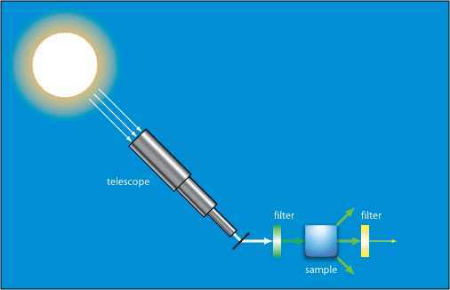
Raman spectroscopy is a tool scientists use to figure out what something is made of by using light — without damaging the object.
Just like light helps us see the world around us, it can also help us see what’s inside things. In Raman spectroscopy, a laser (a very focused beam of light) is shined on a material. Most of the light bounces back the same way — but a tiny bit changes because it interacts with the molecules inside.
That small change in the light gives scientists a sort of “chemical fingerprint” that tells them what the material is made of. It’s a clever way to study things without touching or breaking them — just by watching how light behaves.
Imagine you’re a detective, but instead of using magnifying glasses or fingerprints, your special tool is light.
One day, you get called to investigate different materials — water, silicon, carbon, and even some biological stuff like proteins.
You bring your laser flashlight with you. But this is no ordinary flashlight — it shines a very specific color of light, called a wavelength. Depending on what you want to study, you pick a different color or imagine color with something like vibe.
Case 1: Water (H₂O)
You shine your green laser light (532 nm wavelength) onto a drop of water. Most light just bounces off. But some of it changes slightly — this change happens because the laser light “talks” to the tiny bonds between hydrogen and oxygen atoms inside the water, its like they vibe with eachother. That vibe is like a secret code revealing the water’s molecular dance.
Case 2: Silicon (Si)
Next, you visit a silicon wafer, the base of computer chips. You switch your laser to green or red light (532 nm or 633 nm). When the light hits silicon, it interacts with the crystal’s atoms vibrating in a very specific way just like you vibe with your girl. You see a sharp peak in your data at around 520 cm⁻¹ — this is silicon’s signature vibration just like your heart spikes when you see your partner.
Case 3: Carbon materials (like graphite)
Then, you look at some black carbon material, graphite. You use a near-infrared laser at 785 nm to avoid burning the sample. Here, the light reveals two important “notes” — the “D band” at about 1350 cm⁻¹ and the “G band” at 1580 cm⁻¹. These correspond to how carbon atoms are arranged and how perfect or messy the graphite’s structure is.
Case 4: Biological molecules (proteins, DNA)
Finally, you investigate a biological sample — proteins or DNA — shining your laser at 532 nm or 785 nm. The light interacts with bonds like C=O and C-N, producing a unique pattern in the scattered light. This helps you understand the molecule’s structure or if something is changing, like detecting disease markers.
The Big Idea:
In every case, you’re shining light on a material and listening carefully to how the light’s energy shifts after bouncing off molecules. Those tiny changes are like fingerprints — each molecule has its own unique way of dancing with light.
By picking the right laser color and analyzing these shifts, you can identify materials, study their structure, and even detect hidden details without breaking or touching them.
Now let us look at an emerging new field known as Confocal Raman Spectroscopy
🎯 Confocal Raman Spectroscopy: The Deep Vibe Check
In regular Raman spectroscopy, you’re shining light and catching the vibes from the whole crowd — all the molecules talking at once. But what if you only want to hear one voice in the room, even if it’s deep inside?
That’s where confocal Raman comes in — it lets you zoom in, block out all the noise, and focus on just one exact spot, like tuning into a single heartbeat in a packed concert.
Case 1: Water (H₂O) – Zooming in on one drop
With confocal Raman, you’re not just pointing your laser at the puddle — you’re zooming into one tiny part of that water droplet.
You’re saying, “I don’t want the surface chatter, I want to know how the hydrogen and oxygen are vibing deep inside.”
It’s like ignoring the crowd and locking eyes with one person — their molecular dance becomes crystal clear.
Case 2: Silicon (Si) – The focused heart spike
Instead of scanning the entire silicon wafer, you now focus your laser on a precise depth, maybe a specific layer inside a microchip.
You still get that signature 520 cm⁻¹ spike — but now you know exactly where it’s coming from.
It’s not just “I feel the vibe”, it’s “I feel your vibe, right here, right now, and no one else’s.”
Like when you’re deep in a convo with your girl, tuning out the whole world — just that one frequency between you two.
Case 3: Carbon (Graphite) – Layer by layer playlist
Carbon’s got layers — messy ones and perfect ones.
With confocal Raman, you’re not just scanning the surface and getting a blend of D and G bands.
You’re scrolling through the playlist one song at a time.
You can say, “This D band is coming from 3 microns below the surface — and it’s rough down there.”
You can literally build a 3D map of how the structure changes — like exploring someone’s emotional history, layer by layer.
Case 4: Biological Molecules (Proteins, DNA) – Reading a molecule’s diary
Now you’re deep in a cell — not just looking at the cell, but looking inside it, precisely.
You shine your focused laser on the nucleus, or on one tiny protein strand, ignoring everything around it.
The C=O, C-N bonds light up, telling you their secrets.
It’s like whispering, “I’m not just trying to understand you — I’m trying to understand this exact part of you.”
You’re not reading the whole diary — just the one page that matters today.
The Big Vibe:
Confocal Raman Spectroscopy is the introvert’s dream tool — it blocks out the noise, focuses on the moment, and reveals deep, personal truths one spot at a time. Whether you’re looking for a gold necklace in a packed suitcase or a molecule’s unique signal in a complex sample, confocal Raman helps you say:
👉 “I see you. Just you. And I know exactly where you are.”


